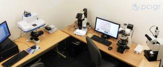
The LaserSpot workstation at the Pacific Centre for Isotopic and Geochemical Research (PCIGR) houses a Raman spectrometer, petrographic and binocular microscopes, and micromill instrument for the preparation, imaging and data processing for both in-situ and microanalysis.
PCIGR’s LaserSpot workstation encompasses a one-stop shop for in-situ and microanalysis. Our Raman spectrometer, petrographic and binocular microscopes, and micromill are connected to computers that are equipped with software for imaging and data evaluation.

(L–R) Horiba XploRA Plus µ-Raman spectrometer, Nikon SMZ18 stereomicroscope, Nikon Eclipse CiPol polarizing petrographic microscope, and New Wave Research micromill in the LaserSpot workstation at PCIGR. Photo: D. Hanano.
Horiba XploRA Plus µ-Raman Spectrometer
The compact Horiba µ-Raman spectrometer enables high-resolution spectral analysis of solid and liquid samples, and is equipped with the following features:
- High-power 532-nm laser
- Full confocal capability for sub-surface analysis
- Flexible slit width, laser filter and confocal hole for optimized tuning on a range of sample materials
- Sensitive CCD detector for high peak-to-background signals
- Notch filter for high-resolution analysis at low wave numbers (<120 cm-1)
- Spectral resolution of 2 cm-1 or smaller for most run settings
At PCIGR, the Horiba Raman spectrometer is typically used for solid- and fluid-phase identification, and structural analysis of minerals and other materials.
Microscopes
LaserSpot has two advanced Nikon microscopes—SMZ18 stereomicroscope and Eclipse Ci POL polarizing petrographic microscope—for high-resolution imaging and petrographic investigation of samples such as mineral separates, mounts, and thick and thin sections.
Both microscopes have these digital imaging capabilities:
- Nikon DS-Fi2 camera
- Nikon DS-U3 digital camera controller for high-definition images (2560 x 1920 pixels)
- PC camera control unit for live image display and editing
New Wave Research Micromill
The New Wave Research micromill enables high-resolution micro-sampling of solid materials for isotopic analyses.
At PCIGR, the micromill is used for analysis where other in-situ micro-sampling approaches (e.g., laser ablation or electron-probe microanalysis) do not generate sufficient quantities of material required for analytical precision.
The micromill is equipped with the following features:
- Drill bits of various thicknesses and shapes for 3-D spatial resolution in the micron range
- Software-controlled motorized stage for exact X, Y and Z coordinates
- Programmable sample traverses and sample maps
- Software to calculate the volume of extracted material
These micromilling capabilities are especially useful for extracting sample material from complex accretionary structures (e.g., skeletal and crystalline material), for analysis via IRMS (δ13C, δ18O), TIMS (Sr isotopes), or multicollector ICP-MS (Nd and Pb isotopes).
Contact
To access the Raman spectrometer, contact Professor Matthijs Smit.
To access other equipment in the LaserSpot workstation, contact Dr. Corey Wall and Dr. Marg Amini.
Back to Laser Ablation and Micromill page